The Charm of Melanogenesis
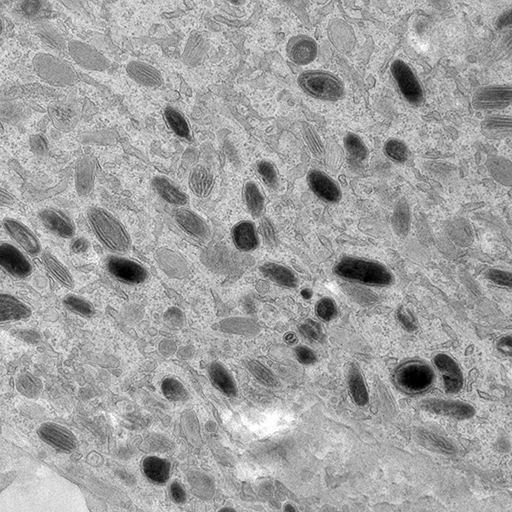
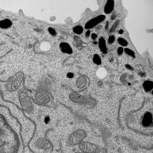
Over the past five years, with the support of the Leo Foundation, our lab has started a new area of research focusing on the intricate cellular processes underlying melanin synthesis. Unexpectedly, our team discovered that a mitochondrial fission factor plays a role in melanosome biogenesis.
Find out more about this fascinating work:
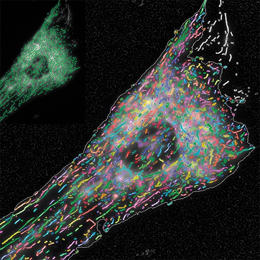
High-Content Imaging and Fluorescence Microscopy
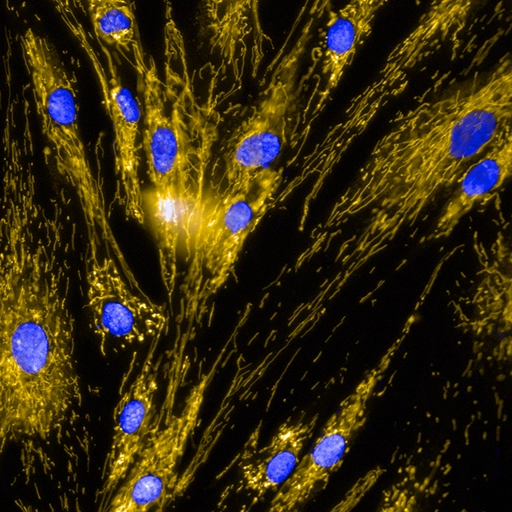
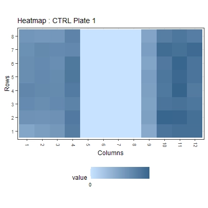
Our lab also focuses on fluorescence microscopy and the development of GFP-based genetically encoded probes to investigate cellular dynamics with high spatial resolution. We have established advanced high-content imaging platforms that enable quantitative and multiparametric analysis of complex cellular phenotypes. Several fluorescence microscopy–based high-content imaging screens are currently running in our laboratory, and we are always enthusiastic about new collaborations and screening projects.
More information about these topics can be found here:
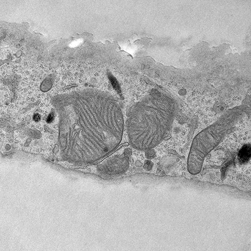
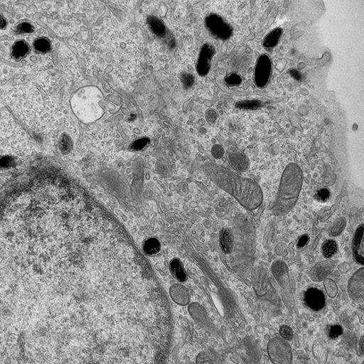
Organelle Contact Sites
Over the past few decades, it has become clear that intracellular organelles communicate with each other in order to meet the cell's physiological needs. This type of communication is mediated by sites of close proximity between organelles, known as 'organelle contact sites'. These sites are extremely dynamic and vary depending on metabolic conditions, stimuli, mitochondrial DNA (mtDNA) and pathological conditions. Our lab is particularly interested in understanding the mechanisms by which cells maintain a certain distance between organelles, either by tethering or spacing biological membranes.
To know more on contact sites:
To know more on mtDNA and contacts:
Funding